Biomolecular Photonics
Prof. Dr. Christoph BISKUP

Prof. Dr. Christoph Biskup
Image: PrivateEmail: christoph.biskup@uni-jena.de
Phone: +49 3641-9-397800
Fax: +49 3641-9-397802
Prof. Biskup is head of the Biomolecular Photonics Group at the Jena University Hospital. He is a member of the board of directors of the Jena Center of Medical Optics and Photonics (CeMOP) and head of the examination committee of the interdisciplinary M.Sc. program Medical Photonics.
One of the aims of the Biomolecular Photonics Group is to establish new photonic techniques and to incorporate them into biologiccal research. The focus is to develop and apply new quantitative multidimensional fluorescence microscopy techniques. Research interests include:
Prof. Biskup is head of the examination committee of the interdisciplinary M.Sc. program Medical Photonics. He gives lectures and courses in:
The laboratories of the Biomolecular Photonics Group offer a wide range of methods used at the interface of physiology, physics and chemistry, including:
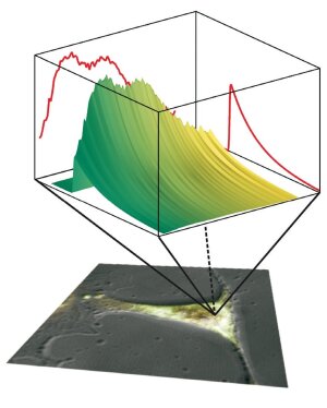
Fluorescence decay surface recorded by using multidimensional fluorescence microscopy techniques. For each voxel of a cell, a time-resolved fluorescence spectrum is recorded.
Illustration: Research group of Christoph BiskupIn conventional fluorescence microscopy, solely information conferred by the fluorescence intensity is used to delineate microscopic structures or perform quantitative measurements by using fluorescent indicator dyes. However, a fluorophore is not only characterized by the intensity of the emitted light, but also by its absorption and emission spectra, by its lifetime in the excited state and by the polarization of the emitted light. The Biomolecular Photonics Group has established several techniques that allow several of these parameters to be recorded simultaneously from a microscopic sample.
Techniques to record time-resolved fluorescence spectra are based on a streak camera system [1] or on the time-correlated single photon counting (TCSPC) technique [2, 3]. Instead of a single fluorescence decay curve, which is obtained by conventional techniques, a fluorescence decay surface can be reconstructed from the data ob-tained by these techniques (see figure).
These techniques can be used to prove the molecular vicinity of proteins by exploiting Förster resonance transfer (FRET). FRET between appropriately labeled proteins occurs only when the labels are in close vicinity (< 10 nm) to each other. Despite a light microscope‘s limited resolution, protein interaction can be visualized in this way [4]. The interaction of key players in cellular signal transduction cascades can be investigated, and the effectiveness of pharmaceuticals to stimulate or inhibit a signal transduction cascade can easily be assessed in high throughput assays.
[1] Biskup et al., Nature Biotechnol. 22, 220 (2004).
[2] Becker et al., Microsc. Res. Tech. 63, 58 (2004).
[3] Biskup et al,., Microsc. Res. Tech. 70, 442 (2007).
[4] Shen et al., Science 315, 1098 (2007).