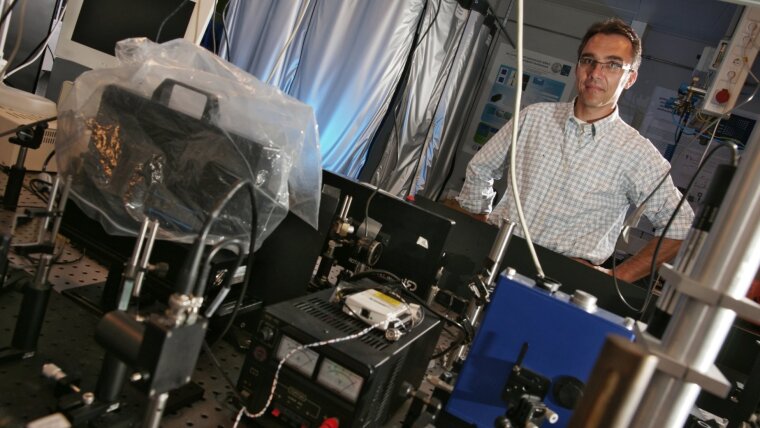
Jürgen Popp
Image: PrivateProf. Dr. Jürgen POPP
Email: juergen.popp@uni-jena.de
Phone: +49 3641-9-48320
Prof. Jürgen Popp is the director of the Institute of Physical Chemistry, the scientific director of the Leibniz Institute of Photonic Technology and a member of the board of directors of the Abbe Center of Photonics. He is the spokesperson of the “Leibniz Center for Photonics in Infection Research” one out of three projects that the German government has put on the national roadmap for research infrastructures. In 2016 he was awarded with the Pittsburgh Spectroscopy Award. 2018 Juergen Popp won the third prize of the Berthold Leibinger Innovationspreis and received the Kaiser-Friedrich-Forschungspreis. In 2019 he was awarded the Ralf-Dahrendorf-Preis für den Europäischen Forschungsraum.
Research Areas
The core research focus of the group of Prof. Popp is biophotonics, i.e. the development and implementation of innovative optical/photonic methods and tools for multiscale spectroscopy and multimodal imaging together with particle- and chip-based molecular point-of-care concepts for biomedical diagnostics. In this context, these biophotonic approaches are utilized and developed according to the needs of pathology, oncology, and infection/ sepsis.
Teaching Fields
Prof. Popp gives courses in:
- Fundamentals of physical chemistry
- Innovative spectroscopy and imaging approaches for biophotonics
- Raman spectroscopy for biomedical diagnostics
Research Methods
The laboratories led by Prof. Popp offer a wide range of laser (micro)spectroscopic methods, at which almost the complete value chain of Raman-based approaches is available:
- Raman (micro)spectroscopy with excitation wavelengths ranging from the UV into the NIR
- Resonance Raman (micro)spectroscopy
- Surface enhanced Raman spectroscopy (SERS)
- IR absorption/ ATR-IR spectroscopy
- Coherent Raman microscopy (coherent anti Stokes Raman scattering (CARS) and stimulated Raman scattering (SRS))
- Second harmonic generation (SHG) microscopy
- Multi-photon excited fluorescence microscopy
- Fluorescence life-time imaging microscopy (FLIM)
- UV/ Vis absorption spectroscopy
- Steady state fluorescence spectroscopy
- Optical coherence tomography
Recent Research Results
Over the last few years, the Biophotonics group has been investigating a multimodal nonlinear imaging approach that has the potential to reliably assess tumour tissue and the success of surgery directly in the operating theatre. The approach combines different nonlinear imaging techniques such as two-photon excited autofluorescence (TPEF), second harmonic generation (SHG) and coherent antiStokes Raman scattering (CARS) [1,2]. For a clinical application this approach was transferred into a compact microscope suitable for clinical use. In order to further extend the applicability of this multimodal microscopy approach for in vivo tissue screening, various endoscopic probe concepts were also realized [3,4,5]. The core of all these setups are robust and alignment-free fiber laser concepts developed by the groups of Prof. Limpert and Prof. Tünnermann in close cooperation with the Biophotonics group [6]. Together with the group of PD Dr. Thomas Bocklitz, innovative image evaluation algorithms for the translation of multimodal images into quantitative diagnostic markers were developed [7,8,9]. Recently, the Popp group was able to show that the realized multimodal imaging approach (I) can be combined with laser tissue ablation for tissue-specific laser surgery and (II) is suitable for the visualization of cold atmospheric plasma-induced tissue alterations for online monitoring of wound or cancer treatment [10,11]. Another focus of the Biophotonics group is an automated high throughput screening of biological cells. The Popp group has developed a high-throughput Raman spectroscopy platform for the automated analysis of cells [12]. This was achieved by completely automating the process chain, i.e. cell detection, Raman spectrum acquisition and chemometric analysis for online cell classification in e.g. multiwell plates or microfluidic environments. Using this automated platform, a large number of cells, e.g. 10,000, can be measured on a time scale of 30 minutes without sample preparation. The applicability of this platform has been demonstrated with several examples, such as (I) differentiated determination of white blood cell counts; (II) identification of circulating tumor cells in a leukocyte mixture; (III) label-free characterization of drug-induced changes [13,14,15].
[1] Chernavskaia et al., Sci. Rep. 6, 29239 (2016).
[2] Heuke et al., Head Neck, 38, 1545 (2016).
[3] Lukic et al.; Optica, 4, 496 (2017).
[4] Zirak et al. APL Photonics, 3, 092409 (2018).
[5] Tragardh et al., Opt. Express, 27, 30055 (2019).
[6] Gottschall et al., TrAC-Trends Anal. Chem. 102, 103 (2018).
[7] Bocklitz et al., BMC Cancer, 16, 534/1-1 (2016).
[8] Bocklitz et al. APL Photonics, 3, 092404 (2018).
[9] Rodner et al. Head Neck, 41, 116 (2019).
[10] Meyer et al., Micromachines, 10, 564 (2019).
[11] Meyer et al., Analyst, 144, 7310 (2019).
[12] Schie et al., Anal. Chem. 90, 2023 (2018).
[13 Mondol et al., Analyst, 144, 6098 (2019).
[14] Mondol et al., Scientifc Reports, 9, 12653 (2019).
[15] Kirchhoff et al., Anal. Chem. 90, 3, 1811 (2018).
