Nanospectroscopy
Prof. Dr. Volker DECKERT

Volker DECKERT
Image: PrivateEmail: volker.deckert@uni-jena.de
Phone: +49 3641-9-48347
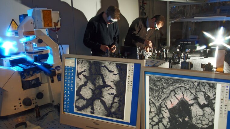
Professor Deckert holds a joint position at the Institute of Physical Chemistry and the Leibniz Institute of Photonic Technology, where he heads the Nanoscopy Department. His main research field deals with investigations of molecular structures within the nanometer range.
Prof. Deckert research interest in general covers fundamentals, method developments, and application of near-field optical spectroscopy. The main target is to use the optical properties of nanoscale metallic structures to reveal the structural conformation and composition of molecules at the highest possible spatial resolution and also understand the underlying principles of high spatial resolution spectroscopy. Research interests include:
Prof. Deckert is involved in the teaching of general physical chemistry and advanced characterization tools.
Prof. Deckert’s laboratories are dedicated to the molecularlevel investigation of surface structures, in particular of biomolecules and heterogeneous catalysis using ultra-high resolution:
Recent experimental evidence made clear that the generally assumed lateral resolution limits in near-field optics have to be revisited. In cooperation with other groups we developed two models to explain tip based nearfield spectroscopy results that demonstrate sub molecular resolution recently. Both models are based on the fact that a plasmonically active nanoparticle can never be a perfect sphere but eventually must show atomic scale features at edges and corners. Such a model is well accepted and used in scanning tunneling microscopy. As for near-field optics, firstly, we could theoretically demonstrate that such features affect the plasmon resonance dramatically in terms of enhancement and field confinement, i.e. lateral resolution. [1] Secondly, if an atomic sized probe is scanning across a molecule, it affects the electronic structure of the molecule depending on the exact location of the probe with respect to the molecule. This so-called chemical effect predicts probe site dependent changes in the vibrational structure of the molecule that are readily detected experimentally as “intensity and spectral position fluctuations” but eventually are related to lateral resolution.[2] Our group also develops new methods to exploit either new information on a specimen, or to improve near-field optical techniques regarding speed or sensitivity. Recent developments are related to laser mode dependent enhancement optimization using a correlated photoinduced force microscopy, a TERS approach and the direct and simultaneous assessment of the tips plasmon resonance and actual temperature directly from a Stokes/anti-Stokes comparison.[3,4] In terms of applications, our main interest is related to bio-related specimen and soft-matter. Based on the experience on bio-spectroscopy with nanometer resolution, the long-term research focus lies on the investigations of dynamic systems, i.e. protein-lipid interactions, immobilized photocatalysts, and drug release of core-shell particles. The latter is a nice example to show how the combined application of different scanning probe techniques provides fast and also structurally specific information about block-copolymer structures used as potential drug carriers.[5] Particularly the correlation of mechanic (force-distance spectroscopy) and near-field Raman (TERS) data allows for a rapid surface precharacterization based on elasticity and adhesion parameters while subsequent specifically targeted TERS experiments result in nanometer resolved structural sensitive information about surface composition, compound mixing, and even unexpected reaction products. This is also a good example of how specific experimental requirements drive the method development and ultimately also the fundamentals of the underlying theoretical concepts.
[1] Trautmann et al., Nanoscale. 9, 391 (2017).
[2] Latorre et al., Nanoscale. 8, 10229 (2016).
[3] Meyer et al., ACS Photonics. 6, 1191 (2019).
[4] Richard-Lacroix et al., Light Sci Appl. 9, 3922 (2020).
[5] Höppener et al., Small. 112, 1907418 (2020).