Single-Molecule Microscopy
Prof. Dr. Michael BÖRSCH
Prof. Dr. Michael Börsch
Image: PrivateEmail: michael.boersch@med.uni-jena.de
Phone: + 49 3641 9-396618
Single-molecule spectroscopy and super-resolution fluorescence imaging of membrane proteins are the topics of our Biophysics Group at the Faculty of Medicine. As a Professor of Microscopy Methods, Michael Börsch has applied single-molecule FRET for nearly 25 years to monitor individual biological nano-machines at work. He is an Adjunct Assistant Professor at SUNY Upstate Medical University (NY, USA) and was a visiting scholar at Stanford University from 2012 to 2014, concentrating on single-molecule research with Prof. W. E. Moerner (Nobel Prize in Chemistry 2014). Prof. Börsch is a member of SPIE and of the Biophysical Society.
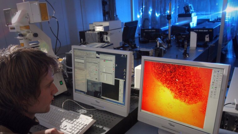
Our Single-molecule Microscopy Group investigates conformational dynamics of single cellular nano-motors, pumps and receptors, for example the enzyme FoF1-ATP synthase. We attach two dye molecules specifically to subunits of these machines and measure their distances continuously within the single protein using Förster resonance energy transfer (FRET). The distance changes manifest the sequences of conformations during either catalysis or transport. Recently, we started super-resolution microscopy with structured illumination (SIM) and single-molecule localization (STORM/ PALM) for the imaging of single bacterial and yeast proteins. Stimulated emission depletion (STED) microscopy with FLIM and homoFRET complements our high-resolution microscopy work.
Our areas of teaching include
We use custom-built confocal microscopes for in vitro single-molecule FRET measurements in solution, equipped with 3D piezo scanners. Lasers provide continuous-wave excitation at 473 nm, 488 nm, 514 nm, 532 nm, 594 nm, 658 nm and 785 nm. Picosecond-pulsed lasers exist from 405 nm to 635 nm, and a super-continuum laser covers the spectrum from 450 nm to 2200 nm. TCSPC electronics count photons simultaneously for up to 6 avalanche photodiodes. Data-acquisition and analysis software was written by our group and includes calculation of fluorescence lifetimes, auto- and cross correlation functions [2], anisotropy and FRET efficiencies at arbitrary time intervals, as well as Hidden Markov Models for analysis of sequential dynamics. One microscope is dedicated for single-molecule FRET and two more for Anti Brownian Electrokinetic trap (ABEL trap) setups [3].
Our biochemical laboratory in Jena is fully equipped to perform cell growth (10-L fermenter system), enzyme purification (FPLC for Ni-NTA, ion exchange and size-exclusion columns, [1]), fluorescence-labeling and ATP-synthase activity measurements [4]. Additional equipment is accessible through central research facilities of the Jena University Hospital (i.e., ultracentrifuges, ultrasonifier systems and fluorescence spectrometers), located in the same building. In our chemistry lab, we produce PDMS microfluidic devices for the ABEL trap in collaboration with the Leibniz Institute of Photonic Technology Jena.
[1] Weiss et al., Nature Materials 17, 89 (2018).
[2] Günther et al., Acc. Chem. Res. 51, 1911 (2018).
[3] Dathé et al., Proc. Spie 10884, 108840N (2019).
[4] Sielaff et al., Molecules 24, 504 (2019).